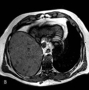
Echogenicity refers to the property of reflecting sound (producing an echo). An echogenic liver is a liver that reflects sound or produces an echo. This has medical relevance only to the medical imaging technique known as an ultrasound.
How Ultrasound Works
Ultrasound is a safe and non-
Ultrasound is safer than many other forms of medical imaging. X-
The downside of ultrasound, though, is that sound is not reflected from substances in the same patterns as light. This means that the patterns formed by echogenicity aren’t exactly the same as those formed by reflected light, so that it can sometimes be more difficult to interpret an ultrasound image than an X-
Nevertheless, many medical problems can be diagnosed using an ultrasound procedure. That’s true of a number of liver conditions, making ultrasound an important diagnostic tool for hepatic illness. The first indicator of liver disease is usually a blood test for hepatic enzymes. Elevation of one or more of these enzymes (of which the liver produces many) can indicate a variety of liver diseases. Medical imaging is normally the next step in diagnosis, with an ultrasound being the least invasive and least risky medical imaging technology available.
Fatty Liver
Fatty liver disease is one of the illnesses that shows up well under ultrasound. Fatty liver consists of excess fatty deposits in the liver. The fat takes the form of lipid globules within the organ. These fat deposits have a higher echogenicity than healthy liver tissue, and so will show up well on an ultrasound test. Fatty liver is not a dangerous condition in itself and usually has no overt symptoms. However, it can progress to more serious liver condition such as cirrhosis of the liver and so is a cause for concern.
Liver Cancer
Cancerous tumors in the liver are also detectable in an ultrasound. The tumor tissue has greater echogenicity than healthy tissue and therefore shows up on an ultrasound. However, it is not possible to tell with certainty whether the abnormal tissue found in an ultrasound is cancerous as opposed to a non-
Fibrosis
Liver fibrosis is a condition in which fibrous growths occur in liver tissue as a result of scarring. This is a stage in the progression of cirrhosis of the liver. Normally fibrosis is diagnosed using a biopsy procedure as is liver cancer. However, the scarring and fibrous growth shows up on an ultrasound as well.
Other Medical Imaging Techniques
Two other medical imaging methods besides ultrasound can provide useful information in diagnosing liver illness. As these methods use different principles than an ultrasound, sometimes they can provide information that does not show up (or does not show up clearly) in an ultrasound.
Computerized tomography (CT) scanning is an X-
Magnetic resonance imaging (MRI) uses a magnetic field to generate information based on the interference or resonance of tissues to the field. MRI can often generate useful information about some liver diseases such as fatty liver. It is less invasive and potentially harmful than CT scanning or other X-
Follow-
When indications of liver disease show up in an ultrasound, further diagnostic procedures are called for to pinpoint the nature and stage of the illness. These procedures can include further blood tests, a liver biopsy, and lifestyle investigation. One important determination is the amount of alcohol the patient consumes. Alcohol abuse is a prime suspect in many liver disorders, but any liver disease with an alcoholic
form also has a non-